Why Plasma Membrane Repair Matters
The Cell’s Most Critical Barrier
Every living cell depends on its plasma membrane to survive. This thin but vital layer separates a cell’s internal environment from the outside world. It controls what enters and exits the cell. Moreover, it allows cells to communicate and cooperate — laying the foundation for complex, multicellular life.
Yet this barrier is fragile. Daily mechanical stress, environmental shifts, and bacterial toxins constantly threaten to puncture it. When damage occurs, cells must act fast. Unrepaired wounds lead directly to cell death. Despite this critical importance, scientists have understood very little about exactly how plasma membrane repair works — until now.
The OIST Study: How Researchers Built the Catalog
A First-of-Its-Kind Protein Catalog
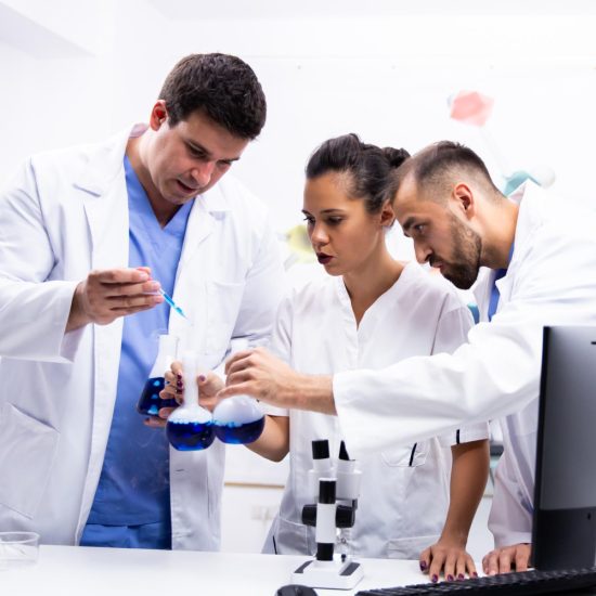
Researchers at the Okinawa Institute of Science and Technology (OIST) have now produced the first large-scale catalog of plasma membrane repair proteins. Their study, published in eLife, used budding yeast as a model organism. In total, the team identified 80 proteins involved in plasma membrane repair. Strikingly, 72 of these had never been reported before.
“This is the first large-scale catalog of plasma membrane repair proteins, and a timeline of how the process unfolds,” said lead author Dr. Yuta Yamazaki of the Membranology Unit at OIST.
The Research Method
The team combined two powerful approaches. First, they ran a proteome-wide screen — scanning thousands of yeast proteins under both normal and stress conditions. Then, they used laser-induced damage to puncture individual cells. This allowed them to track protein movements in real time using advanced live-cell imaging. Together, these methods revealed a precise, step-by-step molecular sequence.
Key Proteins Identified in the Repair Process
How Cells Respond to Damage: A Step-by-Step Sequence
The research uncovered a highly coordinated chain of molecular events. Each stage builds on the last, ensuring the membrane heals quickly and correctly.
Step 1 — Pkc1 Signaling Pathway Activation The first responders are proteins from the Pkc1 signaling pathway. These proteins arrive at the damage site almost immediately. Their rapid arrival triggers the repair response.
Step 2 — Exocytosis Delivers Building Materials Next, exocytosis begins. In this process, vesicles inside the cell fuse with the plasma membrane. They deliver fresh lipids and structural components — essential building blocks for sealing the wound. This stage was expected, as researchers already knew exocytosis played a role in membrane repair.
Step 3 — Clathrin-Mediated Endocytosis Restores Membrane Structure Finally, clathrin-mediated endocytosis (CME) takes over. CME folds the plasma membrane inward, forming a pocket that transports lipids and membrane proteins from outside into the cell. This step likely helps restore the repaired membrane to its normal structure and function.
Bud-Tip Proteins Join the Repair Effort
Another notable finding emerged from tracking protein locations. Many proteins that normally sit at the growing bud tip — where new membrane forms — abandoned their usual posts. Instead, they moved to the damage site to assist with repair.
“The same proteins that create new membranes stop working at the bud and come to repair the damage,” said Dr. Yamazaki. “The machinery of wound repair appears to be similar to the one involved in making new membrane.”
The Surprising Role of Endocytosis
An Ancient Repair Mechanism Uncovered
While Pkc1 activation and exocytosis were anticipated findings, the involvement of clathrin-mediated endocytosis in budding yeast came as a surprise. Scientists had previously observed endocytosis at damage sites in mammalian cells. However, no one had reported it in budding yeast before.
“Endocytosis at the damage site was previously reported in mammalian cells, but not in budding yeast. We show it happens in yeast too,” said Dr. Yamazaki. This finding suggests that CME is an ancient repair mechanism — one that evolved long before mammals and yeast diverged on the tree of life.
Membrane Repair and Human Disease
Why Defects in Repair Cause Serious Illness
Plasma membrane repair is not just a curiosity of cell biology. Mutations in membrane repair proteins directly cause disease. Specifically, defects in these proteins are linked to muscular dystrophy and are associated with accelerated cellular aging. When cells cannot repair their membranes, they die. Over time, widespread cell death contributes to organ-level dysfunction and disease progression.
Furthermore, OIST researchers have previously identified a connection between plasma membrane damage and the aging of cells and organisms. Thus, understanding repair mechanisms has direct implications for longevity research and age-related conditions.
What This Means for Future Research
A Foundation for Human Cell Biology
The catalog produced by this study offers a significant resource for the broader scientific community. Dr. Yamazaki noted: “Our dataset provides a foundation for researchers to investigate plasma membrane repair mechanisms in higher eukaryotes, including human cells.”
In addition, the study opens important questions about evolutionary biology. Because yeast and human cells share these repair mechanisms, researchers can use yeast as a model to explore how membrane repair evolved across cellular life. This, in turn, may yield insights into the deepest origins of cell biology itself.
Ultimately, this foundational research builds the scientific baseline needed to develop future therapies for diseases linked to membrane repair failure — from muscular dystrophy to degenerative conditions associated with aging.