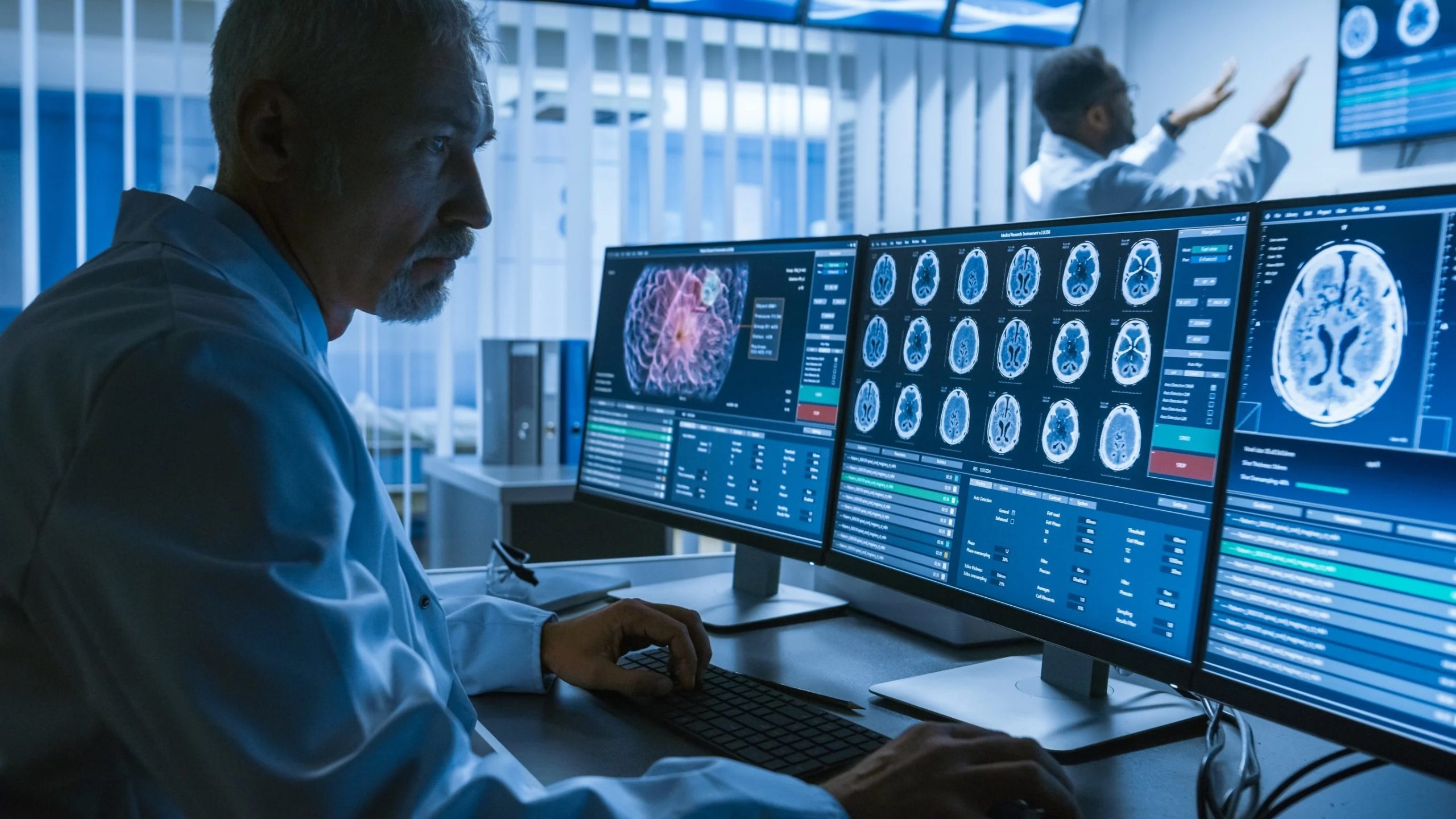
A groundbreaking method developed by the University of Gothenburg bridges the diagnostic gap between CT and MRI scans. Utilizing AI, this innovative software enhances CT images to rival the quality of MRI scans, particularly benefiting dementia diagnosis and neuroradiology. Clinical validation underscores its reliability, reducing false-negative diagnoses and optimizing patient referrals. Its potential extends to diagnosing normal pressure hydrocephalus. The software, trained on over a thousand subjects, offers accurate brain structure measurements. Collaboration with clinics worldwide ensures its integration into healthcare, promising a transformative impact on medical imaging and patient care.
A groundbreaking method is revolutionizing the field of medical imaging by equipping computed tomography (CT) scans with the capacity to yield information comparable to that of magnetic resonance imaging (MRI). This innovative technique, as outlined in a study conducted at the University of Gothenburg, has the potential to significantly augment diagnostic capabilities, particularly in primary care settings, where it can prove invaluable in the diagnosis of conditions like dementia and various other brain disorders.
Computed tomography (CT) has long been favored for its cost-effectiveness and widespread accessibility across healthcare facilities worldwide, including regions with limited access to advanced imaging technologies. However, compared to magnetic resonance imaging (MRI), CT has been regarded as less proficient in depicting subtle structural brain changes and alterations in the ventricular system’s fluid dynamics. Consequently, specific imaging requirements have traditionally been relegated to specialized departments within larger hospitals equipped with MRI technology.
AI-Powered Insights from MRI
The novel software in question serves as a diagnostic aid for radiologists and healthcare professionals entrusted with interpreting CT images. This cutting-edge tool harnesses the power of deep learning, a subset of artificial intelligence (AI), to analyze CT images and generate insights that are akin to those derived from MRI scans of the same subjects.
Michael Schöll, a professor at Sahlgrenska Academy and the lead researcher behind this pioneering study, elaborates, “Our method can extract diagnostically valuable information from routine CT scans, sometimes rivaling the quality of MRI scans conducted in specialized healthcare settings.” The study was a collaborative effort, involving researchers from Karolinska Institutet, the National University of Singapore, and Lund University.
The primary objective is to provide a swift and straightforward method capable of extracting extensive data from routine examinations typically conducted in primary care, as well as select specialist healthcare investigations. Initially, the method focuses on enhancing dementia diagnosis, but its potential applications extend to various facets of neuroradiology.
Reliable Decision Support
The AI-based algorithms driving this innovation have undergone rigorous clinical validation and have the potential to emerge as a swift and dependable form of decision support, significantly reducing the incidence of false-negative diagnoses. Researchers anticipate that this solution will enhance diagnostics in primary care settings, thus streamlining patient referrals to specialist care.
Meera Srikrishna, lead author of the study and a postdoctoral researcher at the University of Gothenburg, emphasizes the significance of this development, stating, “This marks a significant leap forward in the realm of imaging diagnosis. We can now quantify the size of distinct brain structures or regions in a manner akin to the advanced analysis of MRI images. The software enables segmentation of various components of the brain within the image and facilitates volume measurements, even when CT image quality is not as high as that of MRI.”
Expanding the Scope
The software’s training dataset encompassed a total of 1,117 individuals, all of whom underwent both CT and MRI scans. While the current study primarily involved healthy older individuals and patients with different forms of dementia, the research team is actively exploring applications for normal pressure hydrocephalus (NPH).
NPH primarily afflicts the elderly, leading to an accumulation of fluid in the cerebral ventricular system, resulting in neurological symptoms. Approximately two percent of individuals over the age of 65 are affected. Due to its intricate diagnosis and the potential for confusion with other ailments, many NPH cases often go undetected.
Michael Schöll underscores the potential impact of their method in NPH cases: “NPH is a challenging condition to diagnose, and assessing the effectiveness of shunt surgery to alleviate brain fluid buildup can be equally challenging. We believe that our method could significantly enhance patient care in these instances.”
Years of Dedication and a Path Forward
The software’s development spanned several years, and ongoing collaboration with clinics in Sweden, the UK, and the US, alongside a partnership with a healthcare-focused company, is pivotal for the innovation’s approval and eventual integration into healthcare practices. This pioneering technology promises to reshape the landscape of medical imaging and diagnostics, potentially improving patient outcomes and streamlining the healthcare journey for many.

