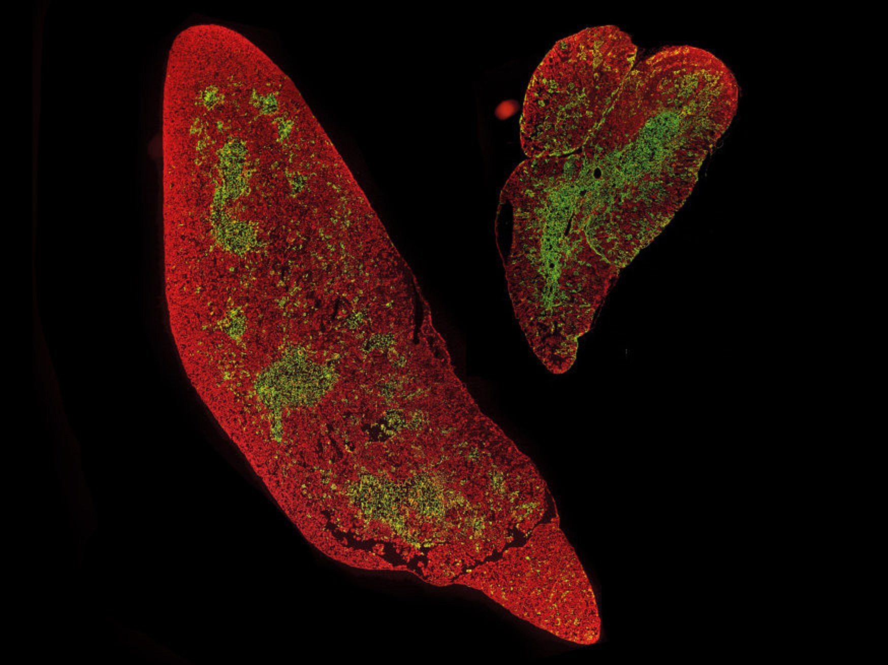
Recent research from Sweden’s Linköping University reveals the thymus, often overlooked in adults, plays a crucial role in the immune system. As the thymus transforms glandular tissue into fat with age, sex, age, and lifestyle influence the pace of this change. CT scans can indicate thymus health, and it affects T-cell regeneration. Lifestyle choices, particularly fiber intake, were linked to thymic fat degeneration. The findings suggest that influencing immune system aging might be possible through lifestyle changes. However, further research is needed to understand the implications of thymus appearance on overall health.
A recent study from Linköping University (LiU) in Sweden suggests that the thymus, a relatively small and lesser-known organ, plays a more significant role in the immune system of adults than previously believed. As individuals age, the glandular tissue in the thymus is gradually replaced by fat, and this process appears to be influenced by factors such as sex, age, and lifestyle. These findings suggest that the appearance of the thymus can serve as an indicator of immune system aging.
Dr. Mårten Sandstedt, MD, PhD, from the Department of Radiology at Linköping University, highlights the newfound importance of assessing the thymus’s appearance in chest CT scans. He points out that this information can offer valuable insights that were previously overlooked.
The thymus, situated in the upper chest, has long been recognized for its role in the development of the immune system in children. However, after puberty, the thymus gradually shrinks and is replaced by fat through a process known as fatty degeneration. This led to the belief that the thymus loses its significance in adulthood. Nevertheless, some animal studies and limited research challenged this notion, suggesting that maintaining an active thymus in adulthood might confer advantages, such as increased resistance to infectious diseases and cancer. Surprisingly, there have been very few studies on the thymus in adults until now.
In the current study, published in Immunity & Ageing, researchers examined the appearance of the thymus in chest CT scans of over 1,000 Swedish individuals aged 50 to 64, participants in the SCAPIS study (Swedish cardiopulmonary bioimage study). SCAPIS encompassed comprehensive health assessments, including lifestyle factors like diet and physical activity, alongside extensive imaging. In this sub-study, the researchers also analyzed immune cells in participants’ blood.
The results revealed considerable variation in thymus appearance, with six out of ten participants displaying complete fatty degeneration of the thymus. This was notably more prevalent in men than women and in individuals with abdominal obesity. Lifestyle choices also played a role, as low fiber intake was associated with thymic fatty degeneration.
This study by Linköping University offers new insights by connecting thymus appearance with lifestyle choices, health factors, and the immune system. The thymus serves as a school for T-cells, a type of immune cell responsible for recognizing foreign invaders like bacteria and viruses while maintaining tolerance to the body’s tissues to prevent autoimmune diseases. The study found that individuals with fatty degeneration of the thymus exhibited lower T-cell regeneration, suggesting that thymus appearance correlates with its functionality.
Professor Lena Jonasson from the Department of Cardiology at Linköping University emphasizes the significance of this association with T-cell regeneration, indicating that CT scans of the thymus reflect not only its appearance but also its function. While factors like age and sex are beyond one’s control, lifestyle-related choices can be influenced, potentially allowing for intervention in the aging of the immune system.
However, further research is necessary to determine whether thymus appearance and immune system aging have implications for overall health. Researchers are now embarking on follow-up studies involving the thymus in all 5,000 participants of SCAPIS Linköping, to assess whether CT scan images of the thymus can predict future disease risk.


