Introduction
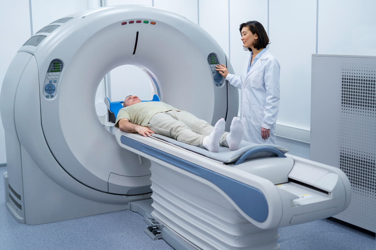
In the ever-evolving landscape of medical imaging, computed tomography (CT) stands out as a cornerstone technology. Since its inception, CT has revolutionized diagnostic capabilities, offering detailed cross-sectional images of the human body. Over the years, advancements in CT technology have propelled the field forward, enhancing image quality, reducing radiation exposure, and expanding clinical applications. This article delves into five of the latest significant trends shaping the scope of computed tomography in medical imaging.
Cardiac CT: Perfusion and Coronary CT Angiography
Perfusion CT
Perfusion CT is instrumental in assessing tissue perfusion, vascular abnormalities, and hemodynamic changes. It plays a crucial role in stroke evaluation, tumor characterization, and assessment of vascular disorders. Advances in dynamic contrast-enhanced CT (DCE-CT) enable real-time assessment of tissue vascularity and pharmacokinetics, holding promise for personalized medicine and treatment monitoring.
Coronary CT Angiography (CCTA)
Coronary CT angiography (CCTA) had a significant presence at the RSNA23 exhibit, where Siemens Healthineers announced the FDA clearance of its Somatom Pro.Pulse, a dual-source CT scanner. This scanner combines the power and speed of dual-source CT technology with embedded artificial intelligence and user assistance features to deliver workflow efficiencies, making it more affordable for a wide range of healthcare facilities.
Photon-Counting CT: A Paradigm Shift
Photon-counting CT represents a paradigm shift in CT detector technology. By directly counting individual photons rather than measuring their energy after interaction with scintillation materials, photon-counting detectors offer superior energy resolution, spatial resolution, and multi-energy capabilities. These detectors facilitate spectral CT imaging, enabling enhanced material decomposition, virtual non-contrast imaging, and improved image quality at reduced radiation doses.
A recent study published in Radiology demonstrated that ultrahigh spatial resolution photon-counting detector CT improved the assessment of coronary artery disease (CAD), allowing for reclassification to a lower disease category in 54% of patients. The study highlights the potential of this technology to improve patient management and reduce unnecessary interventions. However, further validation in real-world comparisons is needed.
AI Integration and Radiomics
The convergence of radiomics and AI is reshaping Computed Tomography imaging analysis. Radiomics extracts quantitative data from medical images to characterize tissue properties and predict clinical outcomes. AI algorithms analyze vast datasets to identify patterns, classify abnormalities, and assist in diagnostic decision-making by detecting abnormalities and quantifying disease burden. Integrating radiomics and AI into CT interpretation enhances diagnostic accuracy, enables predictive modeling, and facilitates personalized medicine approaches and treatment plans.
Dose Reduction Techniques
With increasing concerns about radiation exposure in medical imaging, there is a continued emphasis on developing techniques to reduce radiation dose in CT imaging without compromising diagnostic accuracy. Innovations such as adaptive dose modulation, optimized scanning protocols, and AI-driven dose reduction algorithms are helping to minimize radiation exposure while maintaining image quality and diagnostic confidence.
Portable and Point-of-Care CT
The integration of CT into point-of-care settings is a burgeoning trend, driven by the demand for rapid and accessible diagnostic imaging. Portable and compact CT scanners are being developed for use in emergency departments, intensive care units, and remote healthcare facilities. These systems enable timely imaging, facilitating prompt diagnosis and treatment decisions. Point-of-care CT also holds promise for applications in resource-limited settings and telemedicine.
NeuroLogica’s FDA-approved head-to-toe trauma imaging solution, the BodyTom 64 point-of-care mobile CT scanner, is designed to enhance the user experience and improve clinical workflows through revisions to both the software and the data acquisition system. The scanner has indications for both pediatric and adult imaging and can be used for a variety of needs, bringing the power of innovative imaging to patients’ bedsides safely and efficiently.
Conclusion
Computed tomography continues to evolve, driven by technological innovations and clinical demands. From advancements in image reconstruction algorithms to the integration of photon-counting detectors, the landscape of CT imaging is marked by continuous progress. These trends hold the promise of improving diagnostic accuracy, reducing radiation exposure, and expanding the clinical utility of CT across diverse medical specialties. As we navigate the future of medical imaging, computed tomography remains at the forefront, shaping the way we visualize and understand the human body.
Discover the latest Provider news updates with a single click. Follow DistilINFO HospitalIT and stay ahead with updates. Join our community today!
FAQs
Q1: What is photon-counting Computed Tomography?
A. Photon-counting Computed Tomography is a novel technology that directly counts individual photons, offering superior energy and spatial resolution, and enabling enhanced spectral imaging.
Q2: How does AI integration benefit Computed Tomography imaging?
A. AI integration enhances diagnostic accuracy by analyzing vast datasets to identify patterns, classify abnormalities, and assist in diagnostic decision-making.
Q3: What are the advantages of portable Computed Tomography scanners?
A. Portable CT scanners provide rapid and accessible diagnostic imaging in point-of-care settings, facilitating timely diagnosis and treatment decisions, especially in emergency and remote healthcare facilities.
Q4: How is radiation dose reduction achieved in CT imaging?
A. Radiation dose reduction is achieved through techniques such as adaptive dose modulation, optimized scanning protocols, and AI-driven dose reduction algorithms, ensuring minimal exposure without compromising image quality.
Q5: What is the role of perfusion CT in medical imaging?
A. Perfusion CT is crucial for assessing tissue perfusion, vascular abnormalities, and hemodynamic changes, playing a significant role in stroke evaluation, tumor characterization, and assessment of vascular disorders.


Leave a Reply