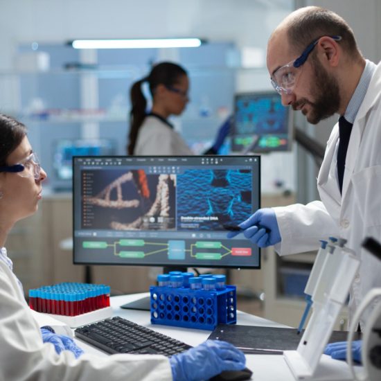
Introduction
Bacteria are far more electrically sophisticated than most people realize. Deep within the microscopic world of electroactive microbes, electrons are constantly being passed from one protein to another — a molecular relay race that allows bacteria to survive in challenging environments and interact with external materials. Now, Cornell University researchers have made a landmark discovery that reveals precisely how this electron relay is organized, and how scientists can control it from the outside.
Published on February 17, 2026, in Nature Communications, the findings shed new light on extracellular electron transfer (EET) — a process with enormous potential for biotechnological applications including microbial energy conversion, bioremediation, and sustainable materials production.
What Is Extracellular Electron Transfer?
Understanding the Basics of Electron Transport in Bacteria
Extracellular electron transfer is the process by which certain bacteria — known as electroactive bacteria — move electrons from inside the cell to outside it, often to external surfaces like minerals or electrodes. This process is essential to how these microbes metabolize energy in environments lacking oxygen.
The challenge lies in the cell’s own architecture. Gram-negative bacteria like Shewanella oneidensis — the most well-known and extensively studied microbe used for electron transport — have multiple structural layers: an inner membrane, an outer membrane, and a periplasmic space sandwiched between them. These layers protect the bacteria against heavy metals, antibiotics, and other environmental threats, but they are largely composed of insulating fatty lipids. In other words, the very structures that protect the bacteria also block the flow of electrons.
Why Electrons Can’t Simply “Swim” Through the Cell
As Cornell’s Peng Chen, the Peter J.W. Debye Professor of Chemistry, explained: “Electrons have to go through the cell envelope: the inner membrane, outer membrane, and the periplasmic space. Electron transfer in biology doesn’t just go through solutions. Electrons do not swim through water. Otherwise, they would get short-circuited.”
This is why proteins are the critical conduits — they act as organized relay stations, guiding electrons safely through each layer of the cell.
The Role of CymA Proteins in Electroactive Bacteria
CymA as the Key Relay Protein
CymA is a critical inner-membrane protein in S. oneidensis that plays a central role in initiating the extracellular electron transfer chain. It passes electrons to downstream protein partners in the periplasm, which then relay them outward through the outer membrane to the external environment.
Despite its known importance, how CymA proteins coordinate their activity with other proteins during this process had remained poorly understood — until now.
Cornell’s Breakthrough Discovery
Biomolecular Condensates: A New Phenomenon in Electroactive Bacteria
Using cutting-edge photoelectrochemistry-fluorescence microscopy, Cornell researchers discovered that during extracellular electron transfer, CymA proteins in the inner membrane do not act independently. Instead, they synchronize and cluster together, forming what scientists call a biomolecular condensate — a concentrated gathering of proteins within a confined region of the membrane.
This condensate formation then drives CymA’s electron-transfer partner proteins in the periplasm to reorganize in the same confined region, creating a highly coordinated, spatially organized relay system.
Biomolecular condensates are a well-documented phenomenon in many types of cells, where they play roles in gene regulation and metabolic reactions. However, this is the first time such condensates have been observed in electroactive bacteria, and the first time they have been linked to electron transfer in any organism.
A Discovery Built on Collaboration
The project was led by Peng Chen and grew from a prior collaboration with co-author Buz Barstow, Ph.D. ’09, assistant professor of biological and environmental engineering at Cornell’s College of Agriculture and Life Sciences. Their earlier work explored how electroactive bacteria interact with semiconducting materials, which inspired Chen to investigate deeper into the mechanics of EET in S. oneidensis. The lead author of the new study is former Cornell postdoctoral researcher Youngchan Park, now an assistant professor at Indiana University.
How Electrochemical Signals Control Protein Patterns
Manipulating Protein Organization from Outside the Cell
One of the most significant aspects of this research is what researchers did after making their discovery: they demonstrated that by applying an external electrochemical signal to the bacteria, they could actively manipulate the spatial arrangement of CymA proteins and trigger condensate formation on demand.
“Many people have applied electrical signals to bacteria,” Chen said, “but we discovered that by applying an electrochemical signal to the cell, it can change the spatial pattern of the protein. The pattern initially is homogeneous, and then you condense it. The electrical signal — basically, the electron transfer — will drive the change of a spatial pattern. That’s a new thing.”
This ability to externally trigger and control protein reorganization at the single-cell level opens the door to a new generation of precision biotechnologies.
Why This Discovery Matters for Biotechnology
Applications in Microbial Energy and Bioremediation
The ability to control extracellular electron transfer with electrochemical signals has far-reaching implications. In microbial energy conversion systems, such as microbial fuel cells, electrons must be shuttled efficiently from bacteria to electrodes. Being able to enhance and control this process could dramatically improve the efficiency of such systems.
Beyond energy, electroactive bacteria are also used in bioremediation — cleaning contaminated environments by breaking down pollutants — and in the synthesis of bioproducts. A better understanding of EET mechanics, combined with the ability to externally manipulate protein organization, provides researchers with a powerful new tool to optimize these applications.
The Research Team and Funding
Co-authors on the study include doctoral student Tianlei Yan, Zhiheng Zhao (Ph.D. ’25), former postdoctoral researchers Bing Fu and Muwen Yang, and Farshid Salimijazi (Ph.D. ’22). The work was supported by the National Institutes of Health (NIH). Imaging was conducted at Cornell Institute of Biotechnology’s Imaging Facility, supported by the New York State Stem Cell Science program and NIH.
Conclusion
Cornell’s discovery that CymA proteins form biomolecular condensates during extracellular electron transfer — and that this process can be triggered by external electrochemical signals — represents a significant leap forward in our understanding of how electroactive bacteria function at the molecular level. As scientists continue to explore ways to harness microbial electricity for energy, environmental, and industrial purposes, this research provides a crucial new foundation for the next generation of biotechnological innovation.