Routine patient photos may reveal how fast the body is aging — and how that pace connects to cancer survival outcomes.
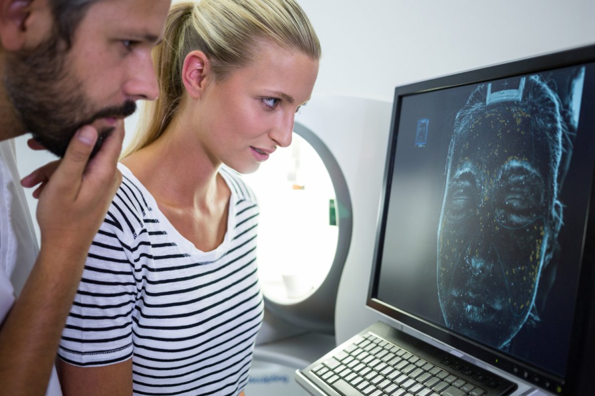
A standard patient ID photo is not usually a medical test. Yet researchers at Mass General Brigham have turned routine facial photographs into a powerful cancer prognostic tool. Their AI-powered system measures how quickly a patient’s face ages over time. Strikingly, that rate of change links directly to survival outcomes.
The study, published in Nature Communications, analyzed data from 2,276 cancer patients. All of them underwent at least two courses of radiation therapy at Brigham and Women’s Hospital between 2012 and 2023.
How FaceAge Reads Biological Age
The AI Behind the Tool
The research builds on FaceAge, an artificial intelligence tool developed by the same team. FaceAge estimates a person’s biological age from a single facial photograph. Earlier findings showed that cancer patients often appeared about five years older than their chronological age. Moreover, higher FaceAge estimates correlated with poorer survival after treatment.
This new study went a step further. Instead of measuring how old someone looked at a single point, researchers measured how fast that apparent age changed between two images.
Calculating Face Aging Rate
The team introduced a metric called Face Aging Rate (FAR). To calculate it, researchers compared two photos from each patient — one from an earlier treatment point and one from a later visit. They measured the FaceAge change and divided it by the time between the photographs.
- A FAR above 1 indicated accelerated aging.
- A FAR below 1 pointed to slower aging.
Additionally, researchers tracked FaceAge Deviation (FAD), which measures how much older or younger a patient appears compared to their actual age in a single image.
When a Face Becomes a Biomarker
Study Cohort at a Glance
The median age at the first radiation therapy course was 63.4 years. Patients ranged from 20.1 to 97.0 years old. Women comprised 50.5% of the cohort and men made up 49.5%. The median time between the two photographs was 286 days, while median follow-up was 35.7 months.
A key finding stood out immediately. Across the entire cohort, median FAR results showed that patients’ facial aging outpaced their chronological aging by 40%. That is a significant biological gap — and it carried measurable consequences.
Faster Facial Aging, Shorter Survival
Time Intervals Reveal a Clear Pattern
Researchers split patients into three groups based on the interval between photos:
- Short-term: 10 to 365 days
- Mid-term: 366 to 730 days
- Long-term: 731 to 1,460 days
The survival differences across these groups were striking. In the short-term group, patients with high FAR had a median survival of 4.1 months, compared with 6.5 months for those with lower FAR. In the mid-term group, median survival was 6.4 months for faster-aging patients versus 12.5 months for slower-aging ones. Furthermore, in the long-term group, the gap widened dramatically: 15.2 months versus 36.5 months.
Hazard Ratios Confirm the Risk
High FAR also carried a significantly elevated mortality risk across all three time windows. In the long-term group, a FAR above 1 carried a hazard ratio of 1.60. After adjusting for variables like sex, race, and cancer diagnosis, the adjusted hazard ratio rose to 1.65 — confirming the association was not a statistical fluke.
Notably, chronological age itself showed no significant link to survival in any time-interval group. FAR, by contrast, consistently predicted outcomes.
Why Rate of Change Matters More
A Dynamic Signal Beats a Snapshot
One of the most important findings involved comparing FAR directly with the one-time FAD measure. Patients with both high FAD and high FAR faced the worst survival outlook. However, over longer intervals, FAR proved to be the stronger predictor of the two.
This suggests that tracking change over time delivers more prognostic value than any single image. The authors noted this mirrors patterns in other medical fields — from blood pressure variability in cardiovascular care to PSA velocity in prostate cancer monitoring.
“Deriving a Face Aging Rate from multiple, routine facial photographs allows for near real-time tracking of an individual’s health,” said Dr. Raymond Mak, a radiation oncologist and co-senior author of the study. “Measuring FaceAge over time may refine personalized treatment planning and help guide the frequency and intensity of follow-up in oncology.”
What the Face May Be Detecting
The authors argue that faster facial aging likely reflects underlying biological strain. This includes cellular senescence, DNA damage, and reduced tissue repair — all processes linked to both aging and cancer progression. In other words, the face acts as a proxy for deeper biological changes that are harder to measure directly.
A related study in The Lancet Digital Health, covering more than 24,500 cancer patients, reinforced this idea. Patients whose FaceAge was at least 10 years older than their actual age showed worse survival. Those within five years of their actual age fared better.
Limits of the Study
Important Caveats to Consider
The findings are promising, but they come with clear constraints. First, the patient cohort was predominantly White (85.1%), which limits how well results generalize across more diverse populations. Facial aging patterns can differ across racial groups, so broader validation is essential.
Second, photo quality, lighting, and facial expression could affect AI performance. Researchers also lacked detailed data on disease progression, cachexia, and treatment toxicity — all of which could influence both appearance and survival.
Third, the photos were not taken at regular study intervals. They were captured at specific radiation therapy time points, which means short-, mid-, and long-term groups may reflect different clinical situations rather than a uniform timeline. Finally, these models have not yet been tested in prospective clinical trials.
What This Means for Cancer Care
A New Layer in Oncology Decision-Making
Currently, FAR is not a stand-alone clinical decision tool. Still, it may add a valuable new layer to cancer care — especially when clinicians need to assess how a patient tolerates treatment over time.
In practical terms, a high FAR might help identify patients better suited for less aggressive palliative treatment rather than escalation. It could also improve risk stratification and support more tailored follow-up schedules.
“Tracking FaceAge over time from simple photos offers a non-invasive, cost-effective biomarker with potential to inform individuals of their health,” said co-author Hugo Aerts, director of the Artificial Intelligence in Medicine program at Mass General Brigham.
The research team has also launched a publicly accessible web portal where individuals can submit facial photographs to receive a FaceAge assessment and contribute to ongoing research. Whether FAR eventually enters routine clinical use will depend on careful validation, equitable performance across populations, and clear proof that the information improves real patient decisions.